Regenerative medicine researchers recreate eggs in vitro in rat model and have moved a promising step closer to helping infertile, premenopausal women produce enough eggs to become pregnant.
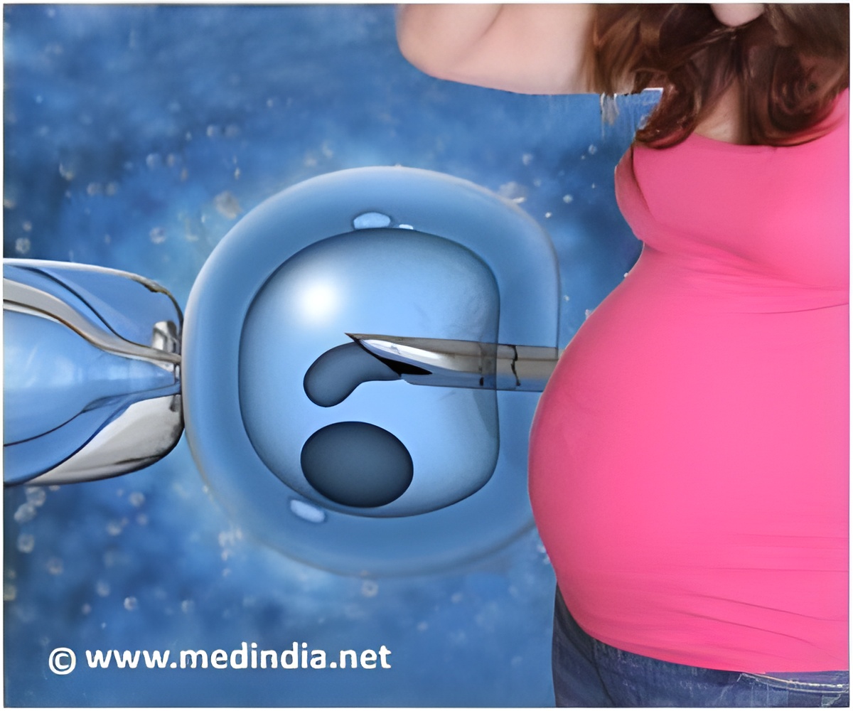
"While conventional hormone replacement therapy is able to maintain female sexual characteristics, it's unable to restore ovarian tissue function, which includes the production of eggs," the study's authors reported. Ovarian tissue function is critical for premenopausal women who desire to conceive.
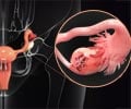
Several fertility disorders can leave premenopausal women without an adequate amount of eggs. These disorders can also prevent a woman's ovaries from secreting enough of the hormones that stimulate egg production. Events such as ovarian operations, an injury, or radiation therapy for cancer can interfere with ovarian function, according to Anthony Atala, MD, FACS, director of the Wake Forest Institute for Regenerative Medicine and chair of the department of urology at the Wake Forest Baptist Medical Center.
Although the causes may vary, about 10 percent of childbearing-age women struggle with infertility,* meaning that these women try for at least one year but are not able to conceive. The U.S. Centers for Disease Control and Prevention says that the most common cause of infertility in premenopausal women is polycystic ovarian syndrome*—an imbalance of sex hormones. This disorder causes irregular ovulation and higher levels of male hormones in affected women.
According to Dr. Atala, the goal of this study was to spur the ovaries to produce the female sex hormones estrogen and progesterone as well as stimulate egg production. The surgeons extracted ovarian cells from three-week old female rats, which would be equiva-lent to about 25 years old in humans. The cells were isolated in a culture of nutrient-dense growth factors for one week. Next, the cells were placed under a collagen gel that allows them to grow three dimensionally instead of in a single layer. The researchers then assessed cell growth, hormone production, and gene expression in the specimens.
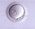
In their early observations, the surgeons found immature oocytes protruding from clusters of ovarian cells. To help the oocytes mature, the surgeons developed a microwell system to keep oocytes inside clusters of ovarian cells. In humans, primordial germ cells or oogonium are the first stage of development into ovums, or mature eggs. The researchers also found that the cells expressed germ cell markers consistent with those of early stage eggs. They observed that the oocytes began to develop zona pellucida, a membrane that forms around an ovum as it develops, and showed a capacity to produce steroids similar to those produced by early stage eggs or follicles.
Advertisement
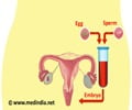
Dr. Atala and his colleagues believe that the newly generated oocytes would be able to mature to a certain stage in humans. The oocytes would then be put back into the female patient to go through natural ovulation and conception, or the oocytes would be fertilized in vitro and then implanted in the uterus. Dr. Atala said because ovarian cell function is restored, a woman using this procedure may be able to produce the necessary hormones and would not need addi-tional hormone replacement therapy.
Advertisement
Source-Eurekalert

