A micro putter has been used by Nagoya University researchers to test the stickiness of individual yeast cells placed on a variety of surfaces.
According to the "cell adhesion model", the more a cell sticks the greater number of chemical bonds it has on its surface. A dead cell would have fewer chemical bonds on its surface so would therefore stick less than a living cell.
The research team has provided compelling evidence that this theory is correct, showing that a cell's adhesion is decreased by more than half once it's died.
Compared to conventional cell testing whereby averages are obtained by staining dead and living colonies with special dyes, the micro-putter could provide a fast and simple approach to testing individual cells and provide a more precise understanding of the biological processes that occur.
Co-author, Yajing Shen, said, "The identification of cell viability is very important in the biological and medical field. Take disease therapy for example: cell viability measurements could be used to evaluate the death of cancerous cells or evaluate cell damage due to toxins."
To fabricate the intricate details of the micro putter, charged particles were fired at a cantilever, using a technique known as focused ion beam (FIB), to remove material from its surface.
Advertisement
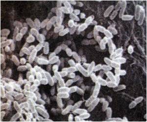
Once inside the ESEM, the yeast cells were delicately placed on three surfaces of varying cell adhesion – tungsten, gold and indium tin oxide – and nudged with the micro putter until they were removed from their spot. The microscope was able to capture images of the movement of the cells whilst the tiny deflection of the putter was recorded as a measure of the cell's adhesion.
Advertisement
"We hope the micro putter can be used to evaluate the actual health conditions of cells in the future. Combined with a single cell surgery technique, the micro putter could help to understand the mechanisms of disease and develop effective new drugs," continued Shen.
Source-Eurekalert


