Duke University researchers have revealed that a protein that controls the metamorphosis of the common fruit fly could help reverse brain injuries.
Incorrect dendrite development or injury has been linked to neurodevelopmental or psychiatric diseases in humans, such as autism, schizophrenia and fragile X syndrome.
Under normal circumstances, neural communication is easy, much like neighbors talking over a fence. But if a neuron is injured or malformed, they frequently don't have the proper dendrites needed to be functional.
"One of the major problems with the nervous system is that it doesn't regenerate very well after injury," said Chay Kuo, M.D., Ph.D., the George W. Brumley assistant professor of cell biology, neurobiology and pediatrics. "Neurons don't multiply, so when they're injured, there's a loss of function. We'd like to know how to get it back."
While prompting such regrowth in the human brain isn't currently possible, dendrite regeneration and arborization -- the branching out of dendrites from the body of the neuron -- are a necessary part of the fruit fly Drosophila's life cycle. In the larval (or worm) state, the fly's nervous system is attuned to what the smooth-skinned worm needs: finding food, locomotion and avoiding attack. As an adult with bristle-covered skin however, the nervous system must be wired for flying, finding mates and laying eggs.
Until now, researchers haven't understood how Drosophila sensory neurons are able to create two separate dendrite branching patterns that successfully serve different kinds of sensory environments, said Kuo, who is also a faculty member with the Duke Institute for Brain Sciences (DIBS). His team set out to find the genetic mechanism that makes it possible. This research, funded by the Alfred P. Sloan Foundation and the George & Jean Brumley, Jr. Endowment, will appear online in the Feb. 27 issue of Cell Reports.
Advertisement
To find out how the drosophila sensory neurons accomplish this change, Kuo's team tagged abdominal sensory neurons with green fluorescent protein (GFP) and followed them through metamorphosis to see if their dendrite branching changed. The dendrite design and architecture was, in fact, different in the adult stage.
Advertisement
Existing literature also pointed Kuo's team to a parallel between the drosophila nervous system and mammals.
"We investigated whether it was possible that Cp1, during metamorphosis, shuttles from the cytoplasm into the nucleus to cleave a transcription factor required for dendrite development, and makes it a new transcription factor for regeneration," Kuo said. "And, that turned out to be true."
The mammalian version of Cp1 is a protein known to be associated with cancer progression and other diseases called lysosomal protein capthesin-L (Ctsl). During the cell cycle, Ctsl can target a transcription factor – a protein that binds specific DNA sequences – called Cut-like 1 (Cux1) that plays a role in gene expression. Ctsl pursues Cux1 inside the nucleus and cleaves it, creating a smaller protein with different transcriptional properties than the original one.
"I feel this discovery is amazing because the major transcription factor involved in how fly sensory neurons grow dendrites in the first place is Cut, and Cut-like 1 is its mammalian homologue," Kuo said. "[Lyons'] initial idea looking into mammalian conservation for answers panned out big. It was serendipity."
By tagging Cut during Drosophila metamorphosis, Kuo's team observed the protein's binding pattern within the nucleus. Before dendrite pruning, Cut binds in big blobs. After the pruning, however, Cut binding is diffused, giving it an opportunity, Kuo said, to bind to different genes during the two dendrite growth phases.
The team translated this finding back to Cp1, discovering that it goes into the neuron nucleus to cleave Cut, making a new transcription factor required for dendrite regeneration after developmental pruning.
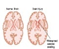
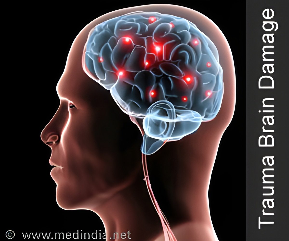
This research could also potentially impact how science and healthcare think about and treat brain injuries, Kuo said. Currently, damaged neurons that have lost their dendrites are unable to properly communicate with their neighbors, rendering them nonfunctional. The problem could be reversed, he said, by helping neurons modify their original developmental program and regrow new dendrites.
"If we can influence this environmental control that changes the development program, it's possible that we could get neurons to integrate and function better after injury," he said.
Source-Eurekalert

