Medindia LOGIN REGISTER
Medindia

Researchers study Vascular Changes Caused by Deep Brain Stimulation Using Brain MRI

by Kathy Jones on Apr 5 2014 2:18 PM

In recent decades, deep brain stimulation has been widely used to treat patients with movement disorders.
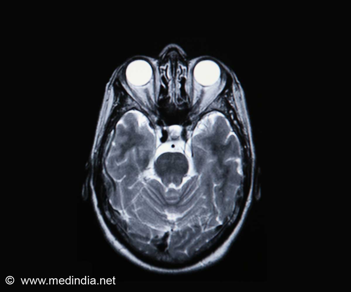
 Researchers study Vascular Changes Caused by Deep Brain Stimulation Using Brain MRI
In recent decades, deep brain stimulation has been widely used to treat patients with movement disorders. Researchers have also paid increasing attention to its use in the treatment of neurological and psychiatric disorders.
However, the influence of subthalamic nucleus or pallidal deep brain stimulation on cerebral vasculature is poorly understood. Even though the metabolic changes caused by deep brain stimulation are being studied using positron emission tomography, the structural changes in cerebral areas like the intracerebral vasculature have not yet been evaluated.

Dr. Byeong Sam Choi and colleagues from Haeundae Paik Hospital, Inje University College of Medicine in Korea performed a retrospective analysis on vasculature changes caused by deep brain stimulation in patients with medically intractable idiopathic Parkinson's disease using double-dose gadolinium-enhanced brain MRI. The dimensions of straight sinus, superior sagittal sinus, ipsilateral internal cerebral vein in the thalamic branch and ipsilateral anterior caudate vein were reduced. These findings, published in the Neural Regeneration Research (Vol. 9, No. 3, 2014), demonstrate that bilateral deep brain stimulation of the subthalamic nuclei affects cerebral venous blood flow.


Source-Eurekalert


Advertisement