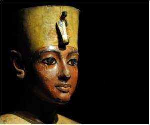
To uncover the story behind one of the earliest surviving Egyptian mummies, a Virginia museum is using CT scan.

The information gathered would help provide greater detail of the body, create a 3-D digital model and even reconstruct the face of the mummy that has been on display off and on since being acquired by the museum in 1953, Fox News reported.
Little is known about Tjeby, who was buried in a rock-cut tomb at a site known as Sheikh Farag in upper Egypt and excavated in 1923.
What museum officials do know is that he dates to between 2150 and 2030 BCE, a time of instability in Egypt, with the breakdown of central authority and economic decline. Previous research suggests Tjeby was 25 to 40 years old when he died.
Experts hope a closer look at data will help piece together more biographical information, such as Tjeby's specific age, diet and cause of death.
They also will look at the materials used to mummify the body and the amount of soft tissue that has survived, and will determine whether organs have been removed, as they were in mummies from later periods.
Advertisement


