Using MRI and a technique-driven by the visual effects industry, the world's most realistic model of the human hand's musculoskeletal system in motion is developed.

‘Combined visual effects techniques and medical imaging create a precise model of the human hand in motion.’


"The hand is very complicated, but prior to this work, nobody had built a precise computational model for how anatomical structures inside the hand actually move as it is articulated," said study co-author Jernej Barbic, an Andrew and Erna Viterbi Early Career Chair and Associate Professor of Computer Science.Designing better prosthetics
To tackle this problem, Barbic, computer animation and physically-based simulation expert, and his Ph.D. student, Bohan Wang, the study's lead author, teamed up with George Matcuk, MD, an associate professor of clinical radiology at Keck School of Medicine of USC. The result: the most precise anatomically based model of the hand in motion.
"This is currently the most accurate hand animation model available and the first to combine laser scanning of the hand's surface features and to incorporate an underlying bone rigging model based on MRI," said Matcuk.
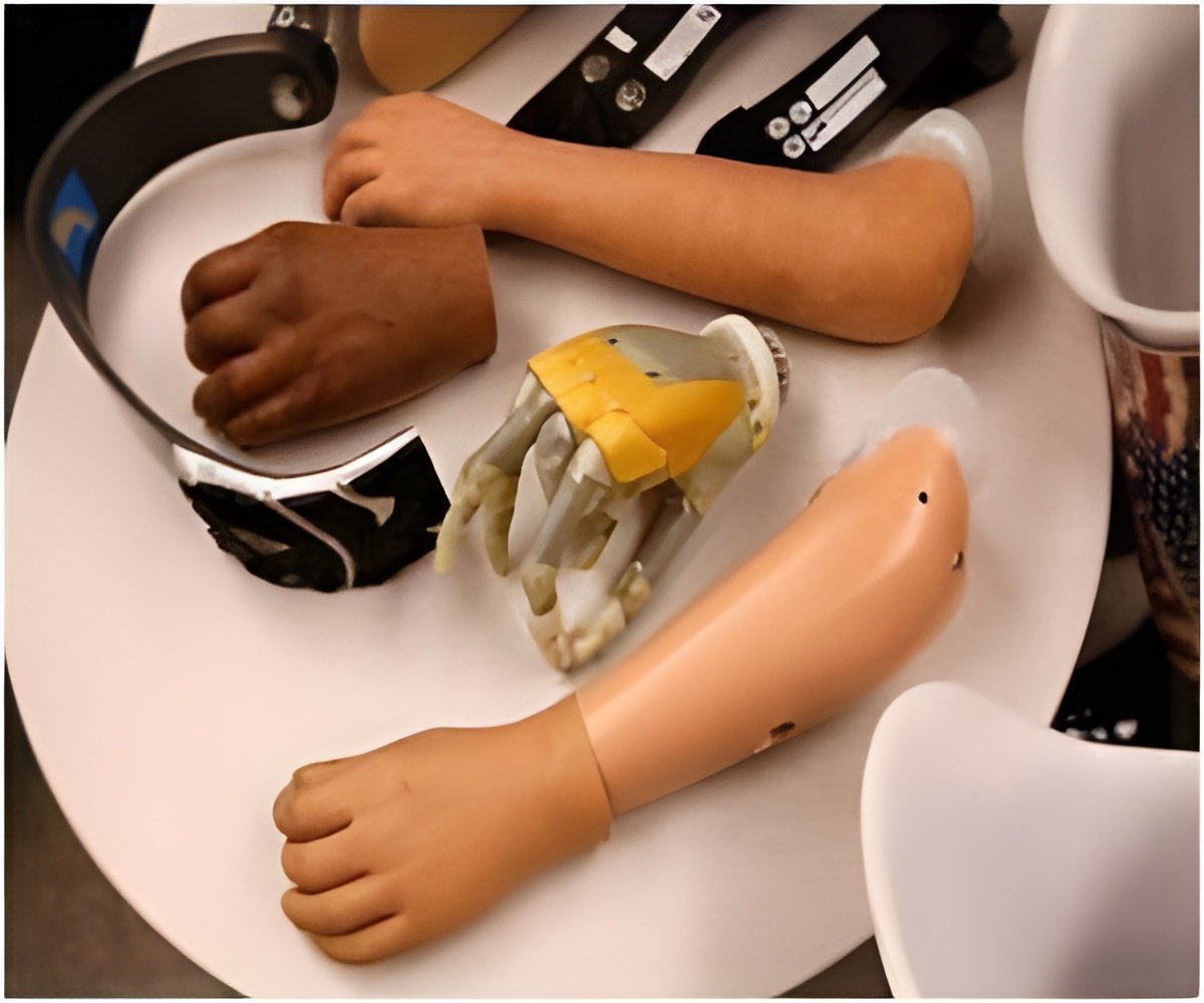
In addition to creating more realistic hands for computer games and CGI movies, where hands are often exposed, this system could also be used in prosthetics, to design better fingers and hand prostheses. "Understanding the motion of internal hand anatomy opens the door for biologically-inspired robotic hands that look and behave like real hands," said Barbic.
"In the not-so-distant future, the work may contribute to the development of anatomically realistic hands and improved hand prosthetics."
Advertisement
A long-standing challenge
To improve realism, virtual hands should be modeled similarly to biological hands, which requires building precise anatomical and kinematic models of real human hands. But we still know surprisingly little about how bones and muscles move inside the hand.
Advertisement
"Holding the hand still in a fixed pose for 10 minutes is practically impossible," said Barbic. "A fist is easier to hold steady, but try semi-closing your hand, and you'll find you start to shake after about a minute or two. You can't hold it still for 10 minutes."
To overcome this challenge, the researchers developed a manufacturing process using lifecasting materials from the special effects industry to stabilize the hand during the MRI scanning process. Lifecasting involves making a mold of the human form and then reproducing it in various media, including plastic or silicone.
Barbic, who worked on the Oscar-nominated film The Hobbit: The Desolation of Smaug, landed on the idea after seeing an inexpensive hand-cloning product in a visual effects store in Los Angeles while working on a previous project. "That was the eureka moment," said Barbic, who has long pondered a solution for creating more realistic virtual human hands.
First, the team used the life-casting material to create a plastic replica of the model's hand. This replica captures extremely detailed features, down to individual pores and tiny lines on the hand surface, which were then scanned using a laser scanner.
Then, the lifecasting process was used again, this time on the plastic hand, to create a negative 3D mold of the hand out of a rubber-like elastic material. The mold stabilizes the hand in the required pose. The mold was cut in two parts, and then the subject placed their real hand into the mold for MRI scanning.
"As we refine this work, I think this could be an excellent teaching tool for my students and other doctors who need an understanding of the complex anatomy and biomechanics of the hand." George Matcuk With assistance from radiology expert Matcuk, a practicing medical doctor at USC, the hand was then scanned by the MRI scanner for 10 minutes. This procedure was repeated 12 times, each time in a different hand pose. Two subjects, one male and one female, were captured in this way. Now, for every pose, the researchers knew exactly where the bones, muscles, and tendons were positioned.
After discussing the anatomical features of the MRI scans with Matcuk, Barbic and Wang set to work building a data-driven skeleton kinematic model that captures complex real-world rotations and translations of bones in any pose.
They then added soft tissue simulation, using the finite element method (FEM) to compute the motion of the hand's muscles, tendons and the fat tissue, consistent with the bone motion. This model, combined with surface detail allowed them to create a highly realistic moving hand. The hand can be animated in any motion, even movement which is very different from the captured poses.
Going forward
The team, which recently received a grant from the National Science Foundation to take their work to the next stage, plans to build a public dataset of multi-pose hand MRI scans, for ten subjects over the next three years. This will be the first dataset of its kind and will enable researchers from around the world to simulate better, model, and re-create human hands. The team also plans to integrate the research into education, to train Ph.D. students at USC and for K-12 outreach programs.
"As we refine this work, I think this could be an excellent teaching tool for my students and other doctors who need an understanding of the complex anatomy and biomechanics of the hand," said Matcuk.
The team is currently working on adding better awareness of muscles and tendons into the model and making it real-time. Right now, it takes the computer for about an hour to create a minute-long simulation. Barbic and Wang hope to make the system faster without losing quality.
Source-Eurekalert


