A new study has revealed that human-derived stem cells can spontaneously form the tissue that develops into the part of the eye that allows us to see.
"This is an important milestone for a new generation of regenerative medicine," Yoshiki Sasai, senior author of the study from the RIKEN Center for Developmental Biology, said.
"Our approach opens a new avenue to the use of human stem cell-derived complex tissues for therapy, as well as for other medical studies related to pathogenesis and drug discovery," Sasai said.
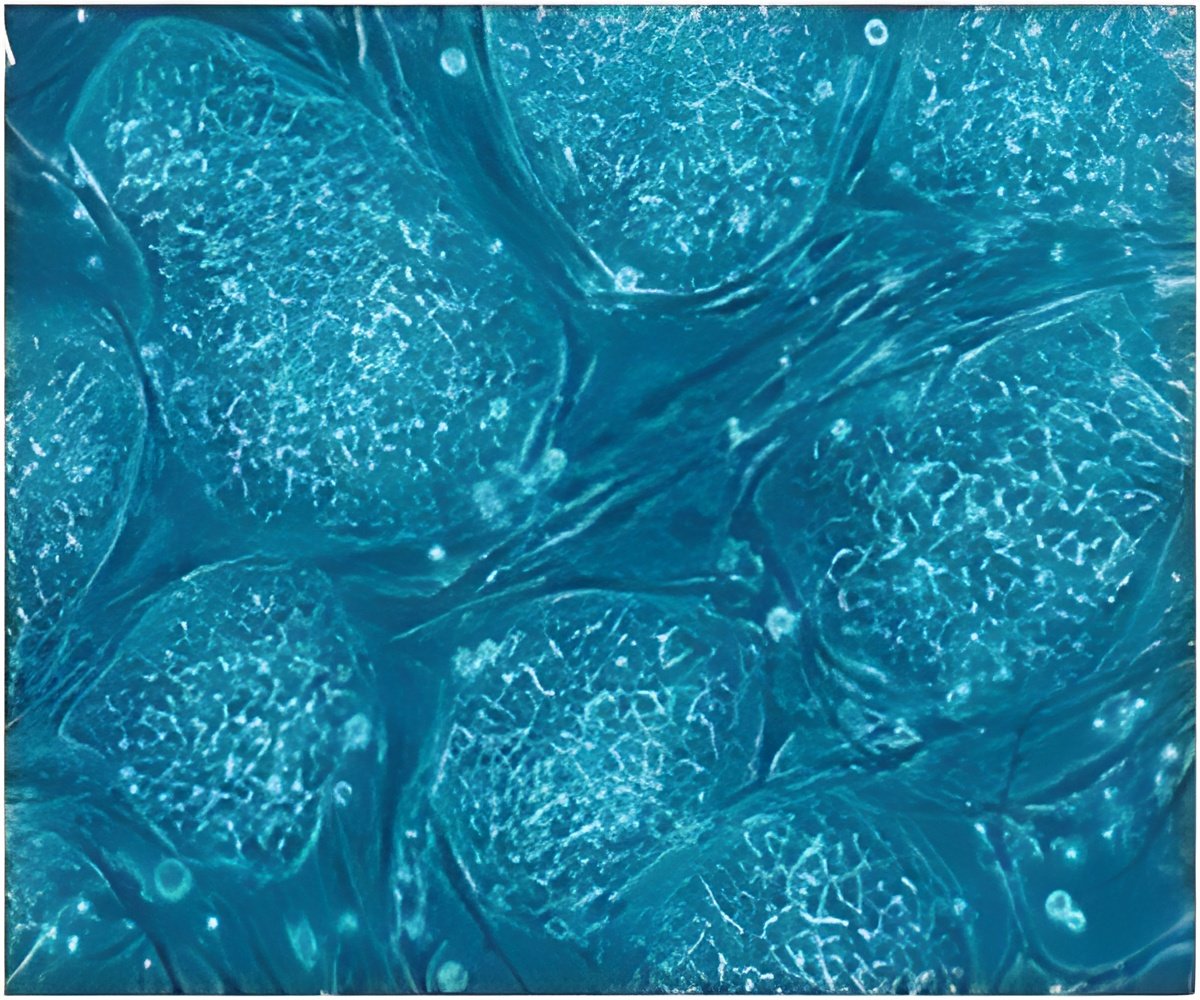
During development, light-sensitive tissue lining the back of the eye, called the retina, forms from a structure known as the optic cup. In the new study, this structure spontaneously emerged from human embryonic stem cells (hESCs)-cells derived from human embryos that are capable of developing into a variety of tissues-thanks to the cell culture methods optimized by Sasai and his team.
The hESC-derived cells formed the correct 3D shape and the two layers of the optic cup, including a layer containing a large number of light-responsive cells called photoreceptors.
Since retinal degeneration primarily results from damage to these cells, the hESC-derived tissue could be ideal transplantation material.
Advertisement
For instance, the hESC-derived optic cup is much larger than the optic cup that Sasai and collaborators previously derived from mouse embryonic stem cells, suggesting that these cells contain innate species-specific instructions for building this eye structure.
Advertisement
The study has been published in the journal Cell Stem Cell.
Source-ANI

