- Anencephaly: Genetic and Rare Diseases Information Center (GARD), National Center for Advancing Translational Sciences (NCATS), National Institutes of Health - (https://rarediseases.info.nih.gov/diseases/5808/anencephaly)
- Anencephaly: St. Louis Children’s Hospital - (http://www.stlouischildrens.org/diseases-conditions/anencephaly)
- Anencephaly: Cleveland Clinic - (https://my.clevelandclinic.org/health/diseases/15032-anencephaly)
- Facts About Anencephaly: Centers for Disease Control and Prevention - (https://www.cdc.gov/ncbddd/birthdefects/anencephaly.html)
What is Anencephaly?
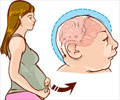
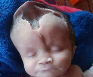
Anencephaly comes from the Greek (An=without + encephalos=brain). Also known as “open skull” this birth defect affects the development of the brain and skull. Thus, a baby is born with an underdeveloped or almost completely absent brain and an incomplete skull and scalp.
Technically speaking, anencephaly is a type of neural tube defect (NTD). The neural tube is a hollow structure which forms the brain and spinal cord. Any defect in the development of the neural tube can result in anencephaly, amongst other congenital abnormalities like spina bifida.
Anencephaly can sometimes result in absence of the entire cerebrum. This part of the brain is responsible for cognitive functions such as memory, judgement, thinking and reasoning. The cerebrum has other functions as well. These include vision, hearing, touch, coordination and movement.
A baby with anencephaly is born blind, deaf, unconscious, and cannot feel anything.
Besides the cerebrum, the cerebellum, which is responsible for fine motor activity, may also be affected. Most babies with anencephaly are stillborn (75%), some die at birth, while some may survive a few hours or at the most a few days.
It should be noted that a baby with anencephaly can be born to a couple who never had any family history of the disease. Importantly, once a baby is born with this birth defect, the chances of future pregnancies having the defect or a related defect such as spina bifida can increase by 4 to 10%.
Epidemiology of Anencephaly
Neural tube defects are a leading cause of birth defects with a global prevalence of 1-2 per 1000 births.. The prevalence of NTD in India varies between 0.5 to 11 per 1000 births. The birth prevalence of NTD including anencephaly varies with geographical location, race, ethnicity, and sex.
Anencephaly occurs in 1 in 5000 births worldwide. It is 2-4 times more prevalent in female fetuses. A wide variation in the frequency of anencephaly has been reported from different parts of India. It ranges approximately between 1.8 to 7 per 1000 live births and is most prevalent in Punjab, Haryana, Rajasthan and Bihar.
What are the Causes of Anencephaly?
It has been found that anencephaly is caused due to multifactorial traits, which involve a combination of genetic and environmental factors that prevent the closure of the neural tube at the head end, which usually closes by the 3rd or 4th week of pregnancy. How the genetic and environmental factors interact to cause the birth defect is still poorly understood. Some of these factors are briefly discussed below:
- Genetic Factors: Differential expression of several genes have been identified as a cause of anencephaly. The most intensely studied gene is the MTHFR gene. This gene encodes an enzyme called methylenetetrahydrofolate reductase (MTHFR), which processes amino acids that constitute proteins. MTHFR is important for carrying out various biochemical reactions of the vitamin folate (also called vitamin B9). It is also believed that other genes may also influence the processing of folate and therefore hinder the proper development of the neural tube.
- Environmental Factors: As is apparent from the above discussion, folate deficiency is the major environmental factor that affects gene expression, which influences its biochemical processing. Therefore, deficiency of this vitamin is one of the major environmental risk factors for the occurrence of NTD.
- Other Risk Factors: Other risk factors include obesity, diabetes mellitus, excessive heat exposure, and certain anti-epileptic medicines. Besides these, socioeconomic status, educational status, and maternal age are also additional risk factors for anencephaly.
What are the Symptoms & Signs of Anencephaly?
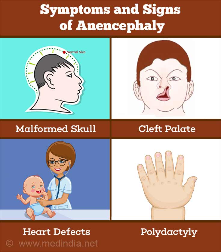
There are several symptoms and signs of anencephaly, which are briefly highlighted below:
- Malformed Skull: The cranial bones can be malformed and a major part of the skull can be missing. This includes the parts covering the front portion (telencephalon) of the brain, consisting of the cerebral hemispheres, as well as the back and sides of the brain.
- Defective Ears: The ears may be defective, often appearing to be folded.
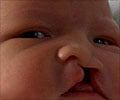
- Cleft Palate: In cleft palate, the top portion of the child’s mouth remains open, which can extend up to the nasal cavity. As a result, the child produces nasalized sounds while speaking.
- Heart Defects: Congenital heart defects may be present as a result of genetic defects.
- Bardet-Biedl Syndrome (BBS): This syndrome can affect anencephalic babies. BBS is a ciliopathic genetic disorder, arising due to primary ciliary dyskinesia that affects many parts of the body. It causes loss of vision due to retinal degeneration (retinitis pigmentosa), extra digits on fingers/toes (polydactyly), obesity, malformed genitals and infertility (hypogonadism), polycystic kidney disease, as well as many other symptoms.
- Alström Syndrome (AS): This syndrome can be present in anencephalic babies. The syndrome is an inherited disorder that affects many organ systems. There is loss of vision and hearing, heart disease (dilated cardiomyopathy), type-2 diabetes mellitus, polycystic liver disease, as well as kidney failure and lung problems.
- Meckel-Gruber Syndrome: This is a rare ciliopathic genetic syndrome that occurs due to primary ciliary dyskinesia and is usually fatal. This disorder is characterized by enlarged kidneys with fluid-filled cysts (polycystic kidney disease), protrusion of the brain at the back of the head (occipital encephalocele), liver fibrosis, and polydactyly.
How do you Diagnose Anencephaly?
A diagnosis of anencephaly can be made before birth, which is technically known as prenatal diagnosis. After birth the features of anencephaly becomes clearly evident upon a physical exam. The baby’s head will appear flat and bones of the skull and scalp will be absent.
The following diagnostic tests are generally performed during pregnancy for evaluation of the baby for anencephaly:
- Alpha-fetoprotein (AFP): This is a blood test that detects AFP, which is a protein produced by the immature liver cells of the fetus and is excreted into the amniotic fluid. Abnormal levels of AFP may indicate brain or spinal cord defects, multiple fetuses, a miscalculated due date, or chromosomal disorders. This test can identify 80-90% of babies having a NTD.
- Amniocentesis: Amniocentesis is performed to determine chromosomal and genetic disorders and certain birth defects. The method involves insertion of a needle through the uterine wall into the amniotic sac to obtain an amniotic fluid sample to estimate AFP or acetylcholinesterase (AChE). Importantly, AChE has been found to be a definitive marker of NTD in early pregnancy. Higher-than-normal levels are indicative of NTD, such as anencephaly. This procedure is very reliable and an anencephaly diagnosis is unlikely to be a false positive.
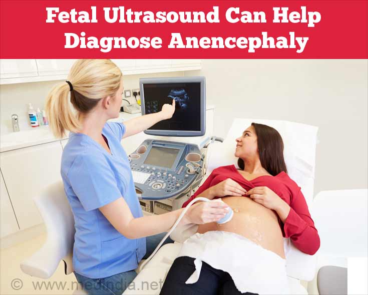
- Fetal Ultrasound: Fetal ultrasound is a diagnostic imaging technique that uses sound waves that are emitted from a transducer and are deflected by the fetus, thereby generating an image on a monitor. Anencephaly is often apparent on an ultrasound by the second trimester.
- Fetal MRI: Magnetic resonance imaging (MRI) is a very sensitive and accurate imaging technique that uses magnetic waves coupled with a computer to generate an image of the internal structures of the body. Fetal MRI can generate very clear images of the developing fetus. The structure of the brain can be seen in greater detail than by ultrasound.
Any of the above procedures can be carried out between the 14th and 18th weeks of pregnancy, while the fetal MRI can be performed at any time during the pregnancy.
What Should you do After a Diagnosis is Made?
The decision of what to do after anencephaly is diagnosed is very difficult to take, since the couples know that the baby will inevitably die. That is why most couples decide to terminate the pregnancy after receiving a prenatal diagnosis of anencephaly. In rare instances, some couples opt to carry on the pregnancy, often based upon religious beliefs, in spite of being fully aware that the baby will not survive.
How do you Treat Anencephaly?
There is no known cure or available treatment regimen for anencephaly. Almost all babies born with anencephaly die shortly after birth, if not already stillborn. As long as the baby is alive after birth, only supportive care can be given.
How do you Prevent Anencephaly?
Supplementation of the diet with folic acid before and during pregnancy can help prevent NTD, such as anencephaly. The standard recommended dose of folic acid is 400 µg daily. Two of the major strategies for preventing anencephaly include the following:
- Food Fortification: Fortification of foods with folic acid and other vitamins can substantially reduce the occurrence NTD such as spina bifida and anencephaly, as is evident from studies conducted in USA and Europe. In the Indian context, staple food such as wheat flour can be fortified to increase the uptake of folic acid in the diet.
- Genetic Counselling: The role of genetic counsellors in preventing the occurrence of NTD like anencephaly is of the utmost importance. Genetic counselling is crucial for reducing the risk of recurrence of anencephaly in a future pregnancy. Advice about taking folic acid, two months prior to conception and throughout the first trimester of pregnancy is recommended for decreasing the recurrence of NTD for couples who have had a previous child with an NTD. Moreover, the need to avoid smoking and drinking alcohol during pregnancy is another important piece of advice that genetic counsellors give to expecting mothers.


