- Pandit HG, BarnettA. Hip and Knee. In Williams NS, Bulstrode C.J.K, O’ Conell PR Bailey and Love’s Short Practice of Surgery 26th edition Taylor and Francis 2013:505
- Hamblen DL, Simpson AHRW eds. Complications of Fractures. In Adam’s Outline of Fractures Including Joint Injuries.12th edition. Churchill Livingstone 2007:52
About
Avascular necrosis or Osteonecrosis is death of a bone due to ischaemia or deficient blood supply. This can have serious consequences like degenerative changes and disabling osteoarthritis of the joint.
The immediate consequence of ischaemia or compromised blood supply to the bone is that the cells die, and if the affected part, as is seen usually, lies within the joint cavity, there is little chance of restoration of blood supply from the surrounding tissues. As a result irreversible changes of structure and form in the bone take place. The normal rigid trabecular structure is lost and the bone becomes gritty and granular. Thus the bone crumbles easily and eventually due to stress because of the muscle tone or body weight it can eventually collapse into an amorphous mass.
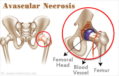
This process can be very slow, taking three to four years or may occur in the year of injury itself. Meanwhile the overlying cartilage also dies as it loses nourishment resulting in the joint undergoing crippling osteoarthritis.
In children, sometimes, the articular cartilage can receive nourishment from the synovial fluid and this allows the deeper layers to survive till the bone is revascularised.
Sites of Avascular Necrosis
The most frequent sites of avascular necrosis are:
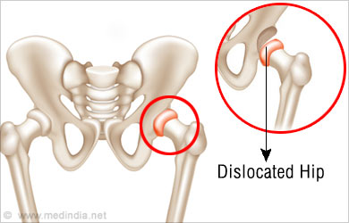
- Head of the femur or the thigh bone after fracture of the femoral neck or dislocation of the hip
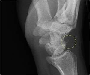
- The scaphoid bone after a fracture of the wrist
- Fractures to the talus bone at the sole of the foot, seen commonly in drivers of vehicles involved in road traffic accidents.
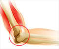
- Dislocated lunate bone at the elbow where the entire bone can undergo avascular necrosis. This occurs when the hand is extended in a fall.
Causes of Avascular Necrosis
- Idiopathic or Unknown
- Secondary to trauma or injury
- Secondary to other pathology like:
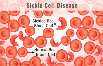
- Heamoglobinopathies
- Caisson s disease
- Hyperlipidemia
- Systemic Lupus Erythematosus
- Gaucher’s disease
- Chronic liver disease
- Antiphospholipid antibody syndrome
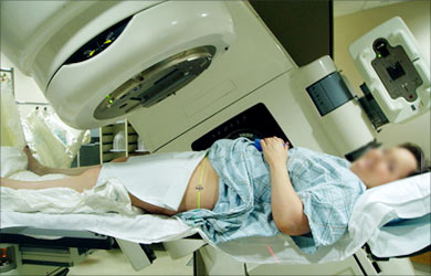
- Radiotherapy
- Chemotherapy
- Human Immunodeficiency virus
- Hypercoagulable states- Protein C and Protein S deficiency
Clinical Features of Avascular Necrosis
In the Femur or Thigh bone:
- Usually occurs in men aged 35-45 years
- Bilateral in over 50% patients
- Frequently asymptomatic in the early stages
- Ache in the groin

On Examination there can be:
- Collection of fluid
- Limp
- Limitation of movement
In the scaphoid bone at the wrist:
- It occurs after fracture to the proximal end of the bone due to fall on an outstretched hand.
- It can go unnoticed
- There can be pain or impairments of wrist movements
- It is a serious complication as it can cause a permanent disability.
Fracture to the talus bone at the sole of the foot can occur after a fall from height. If the bone undergoes displacement, chances of avascular necrosis are very high.
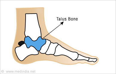
It can be seen in drivers involved in road traffic accidents when the foot pedals cause injury.
Investigations in Avascular Necrosis
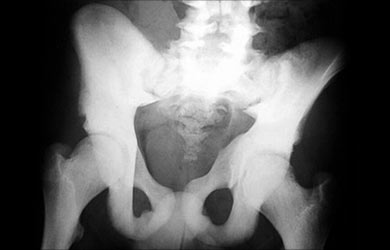
- X Ray of the pelvis
Increased thickening in the first stage followed by resorption of the bone leading to final flattening and collapse of the bone.
- X-ray of the wrist can show thickened, dense appearance when there is avascular necrosis of the scaphoid bone in the wrist.
- X-ray of the foot shows the characteristic dense appearance initially with collapse in the final stages.
- MRI of the bone
Most sensitive and specific investigation
Enables early detection even before the X-ray changes are manifest and also helps to judge the prognosis
Treatment of Avascular Necrosis
Based on the radiological findings Avascular Necrosis is classified into seven stages which are in the precollapse or collapse stage.
Treatment depends on whether it is in the precollapse or the collapse stage
In the femur precollapse group, the principle is to preserve and revascularise the femoral head.
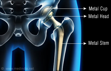
In the collapse group the aim is to replace the femoral head.
Surgical intervention is required as conservative treatment leads to poor results.
In the post collapse stage, femoral osteotomy is done, where the aim is to transfer the weight bearing area and protect the collapsed segment. If degenerative change has set in, it is preferable to consider a replacement.
The scaphoid bone may be amenable to treatment before collapse and development of arthritis.
Bone grafting can be done in specialized hand surgery centres but once arthritic changes have set in, permanent disability of the wrist is to be accepted.
In the foot, surgical correction and excision along with arthrodesis may be required.
Studies are on with regenerative stem cell therapy.
Frequently Asked Questions
1. Who treats Avascular Necrosis?
The orthopaedic surgeon is the specialist who treats this condition.
2. What is Keinbock’s disease?
Idiopathic (of unknown cause) avascular necrosis of the lunate bone in the elbow is called Keinbock’s disease.
3. What is Preiser’s disease?
Idiopathic avascular necrosis of the scaphoid bone in the wrist joint is called Preiser’s disease.
4. What is Aviator’s Astragalus?
This is the name given to fracture of the talus in pilots of crashed aircraft.

