About
The specimen collected from biopsy is processed in one or both of two major ways
a) Histological Section
b) Pathologic Examination
Histological Section:
The sample collected from biopsy technique is made into thin slices and mounted on a glass slide and stained with appropriate staining solution and covered by a glass coverslip for examination.
There are two major techniques for preparation of histologic sections:
- Permanent Section Technique
This technique provides best quality of specimen for examination.
The fresh specimen is immersed in a liquid called a fixative for several hours (the necessary time depends on the size of the specimen).
The fixative, formalin causes the proteins in the cells to denature and become hard and "fixed." Adequate fixation is probably the most important technical aspect of biopsy processing. The fixed specimen is fixed with paraffin wax.
The next morning histologist removes the paraffin- impregnated specimen and "embeds" it in a larger block of molten paraffin. This is allowed to solidify by chilling and is set in a cutting machine, called a microtome. The histotech uses the microtome to cut thin sections of the paraffin block containing the biopsy specimen.
These delicate sections are floated out on a water bath and picked up on a glass slide.
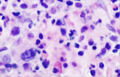
Paraffin wax is dissolved from the tissue block by using solvents. Then the tissues are stained with dyes - Hematoxylin and eosin.
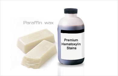
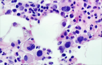
The stain combination, casually referred to by pathologists as "H and E" yields pink, orange, and blue sections that make it easier for us to distinguish different parts of cells. Typically, the nucleus of cells stains dark blue, while the cytoplasm stains pink or orange.
- Frozen Section Technique
This technique allows examining histologic sections within a few minutes of removing the specimen from the patient.
The quality of the tissue sections is not as good as those of the permanent section.
A skilled pathologist and a knowledgeable surgeon can work together to use the frozen section's rapid availability to the patient's great benefit.
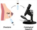
Smears: The specimen is a liquid, or small solid chunks suspended in liquid. This material is smeared on a microscope slide and is either allowed to dry in air or is "fixed" by spraying or immersion in a liquid. The fixed smears are then stained, and examined under the microscope.
Pathologic Examination:
a. The Gross Description
The pathologist first examines the specimen with the naked eye. This is the "gross exam".
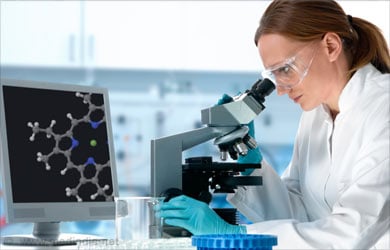
Most biopsies are small, nondescript bits of tissue, so the gross description is useful for identification of the origin of the tissue.
Larger organs removed as biopsies have correspondingly longer and more detailed gross descriptions.
b. The Microscopic Examination
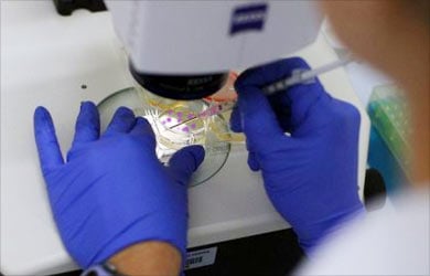
The microscopic description, of the findings is obtained from examination of the glass slides under the microscope.
c. The Diagnosis
The final diagnosis of the biopsy specimen is mentioned in the report. The report explains the name of the organ used in biopsy. The site in the organ where the biopsy is obtained. The type of surgical procedure used in obtaining the biopsy.

