- Boubaker A, Prior JO, Meuwly J and Bischof-Delaloye A. Radionuclide Investigations of the Urinary Tract in the Era of Multimodality Imaging. J Nucl Med 2006; 47 (11): 1819-1836
- Nuclear Medicine - (http://www.nibib.nih.gov/science-education/science-topics/nuclear-medicine)
- Your Kidneys & How They Work - (http://www.niddk.nih.gov/health-information/health-topics/anatomy/kidneys-how-they-work/pages/anatomy.aspx)
What is Isotope / Radionuclide Cystogram?
Radionuclide cystogram or radionuclide cystography is a diagnostic procedure done by nuclear medicine specialists to study the urinary bladder. The procedure uses a radioactive tracer to detect functional and sometimes structural abnormalities of the urinary bladder. This is captured by a gamma camera and the dynamic images of the study can be viewed on the monitor to make a report.
Radioactivity substances used are inert and completely safe; they have a miniscule amount of radiation that is harmless to the body and the ‘term radioactive’ should not worry the parents or the patient having the test. You must remember that in our everyday life, we are exposed to small amount of radiation from the sun and the appliances that we use.

How Does the Urinary Tract Function?
The urinary tract consists of the kidneys, ureters, urinary bladder and urethra. The kidneys filter waste material from the blood and produce urine. The urine passes through two tube-like structures called the ureters into a muscular pouch called the urinary bladder. Once the bladder gets full, the patient gets an urge to pass urine, which is excreted out of the body through the urethra. The urethra is long in males and passes through the penis, while it is short in females.
Why is a Radionuclide Cystography / Isotope Cystography Done?
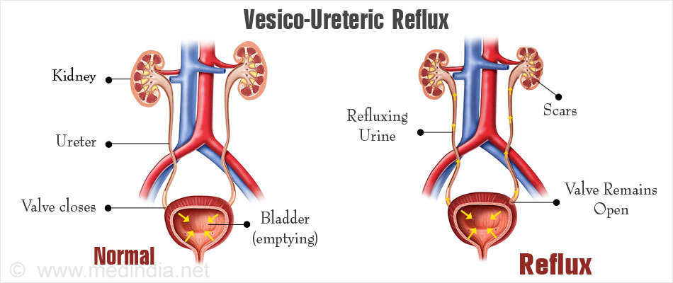
Isotope Cystography or radionuclide cystography is a bladder scan commonly done to diagnose a condition called vesicoureteral reflux. When the normal bladder contracts to pass urine, the urine should pass out through the urethra. However, in some cases, some urine passes back into the ureters and could be responsible for repeated infections. This condition is called vesicoureteral reflux.
Vesicoureteral reflux is sometimes an inherited condition, therefore siblings of a patient may also suffer from the same condition. In other cases, it may be due an obstruction to the flow of the urine at the level of the urethra, for example, due to prostate enlargement in adult males. The patient may develop dilatation of the upper urinary tract. Vesicoureteral reflux is a common cause of urinary tract infections in children. The repeated infections could result in kidney damage if not diagnosed early.

Radionuclide cystography may also be used as a follow-up for patients with vesicoureteral reflux to check if the condition has subsided.
Radionuclide cystography can also detect large structural defects of the bladder. However, it may not be able to pick up smaller bladder defects like diverticula.
What are the Types of Isotope Cystography / Radionuclide Cystography?
During a radionuclide cystography, a small amount of radioactive substance called a radiotracer is either injected into a vein or directly instilled into the bladder. Technetium 99m is a commonly used radioactive tracer to assess the urinary tract. It emits gamma rays, which can be detected using a gamma camera. The gamma camera converts the signals into electric signals that are passed on to a computer to create images of the bladder, which are interpreted by a nuclear medicine specialist.
There are two types of radionuclide cystography:
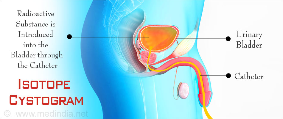
- Direct Radionuclide Cystography: In direct radionuclide cystography, a catheter is placed into the bladder. The radionuclide tracer is instilled into the bladder through the catheter, and images are taken. Images during the filling as well as the emptying of the bladder can be recorded, and bladder dysfunction can be noted.
- Indirect Radionuclide Cystography: Indirect radionuclide cystography is a continuation of the renal scan procedure. The radioactive tracer along with a guiding molecule like mercaptoacetyltriglycine (MAG3) is injected into a vein and images are taken with a gamma camera as it passes through the bladder, and out through the urethra. Since the bladder fills by itself rather than fluid being inserted through the urethra, the bladder can be studied under more physiological conditions. This procedure can be done only in toilet-trained children.
How Should the Patient Prepare for the Procedure?
The radionuclide cystography does not need any preparation or hospitalization. The patient can visit the scanning center on the same day as your appointment.
If the patient is a child, the parents and the nurses have to ensure that the child is as comfortable as possible during the procedure and with their favorite toy if possible. Tests like ultrasound and urine test may be advised before undergoing bladder scan.

When the doctor advises an isotope cystogram, he should be informed if the patient suffers from any allergies or illnesses or takes any medications.
Jewellery should not be worn during the test. If the test is advised in a woman, she should inform the doctor if she is pregnant.
An antibiotic may be prescribed for direct cystography prior to the procedure.
What Happens During a Radionuclide Cystogram?
During a direct procedure, the patient will be asked to change into a gown and to lie down on the scanning table with feet apart. The doctor will pass a small tube through the urethra.
An anesthetic gel may be used to avoid pain during the procedure. The fluid that contains the radionuclide will be instilled into the bladder to fill it.
Once the bladder is full, the patient will be asked to empty the bladder. Scanning will be carried out throughout the procedure of voiding, the procedure being referred to as voiding cystourethrogram.
Scanning after emptying will indicate if the bladder is being emptied completely or not.
An indirect radionuclide cystography follows a renal scan. When the dye passes through the bladder and when the patient voids, scans are obtained with a gamma camera.
How Does the Radiologist Interpret the Results?
Based on the scanned images, the radiologist can come to certain conclusions. For example, In the case of reflux, the radionuclide may move upward from the bladder and may be detected in the ureter. In severe cases of reflux, it may extend to the renal pelvis and even into the kidneys.
If some radionuclide is detected in the bladder even after the patient has passed urine, it indicates incomplete emptying of the bladder. Incomplete emptying or residual urine in the bladder may be a result of an obstruction like an enlarged prostate, or due to a neurological problem that does not allow the bladder to contract properly. Incomplete emptying can result in repeated bladder infections.
How Long Does It Take?
The procedure may take around an hour or two to complete.
What Happens After the Procedure?
Once the scan is complete, the catheter is removed and the patient will be able to go home.
The patient will be asked to drink plenty of water so that the person feels the urge to pass urine frequently, and the radiotracer can be washed out from the body in the urine.
If they have any discomfort like burning in the urinary passage or of they develop fever with shivering, they should report to the doctor or hospital immediately. They maybe prescribed a short course of antibiotics after the test to be taken for a few days to avoid possible urinary infection.

What are the Benefits of This Procedure Over Other Procedures to Detect Vesicoureteral Reflux?
The other procedure done to diagnose vesicoureteral reflux is radiological micturating cystogram. In this procedure, a contrast is filled into the bladder through a catheter. Images are taken during filling and emptying of the bladder as well as after emptying.
A radionuclide cystography has the following advantages over a micturating cystogram:
- Exposure to radiation is less. This is particularly important since the procedure is often carried out in children.
- The test is more sensitive and the results are more reliable.
- More quantitative data is obtained.
What are the Complications and Risks?
A radionuclide scan is usually without complications. However, it should be avoided in pregnant women and should be done with caution in individuals who have an allergy to the tracer or any substance such as antibiotics that are being prescribed. Possible complications include:
- Small risk of radiation to the fetus for pregnant women.
- Urinary tract infection.
- Trauma to the urethra or bladder, due to which the urine may appear slightly pink and the patient may experience some discomfort for a couple of days following the procedure.


