Diagnosis
Testicular cancer can be easily diagnosed in most cases by careful self-examination.
Testicular cancer can be easily diagnosed in most cases by careful self-examination, preferably after a hot water bath, when the scrotal bags are more loosened and relaxed; during this time any lump or change in size or texture can be easily detected. In other cases, a doctor detects the lump during a routine health exam.
Any change in the size, shape or texture of the testicles is a cause for concern. The individual must be subjected to a medical examination.

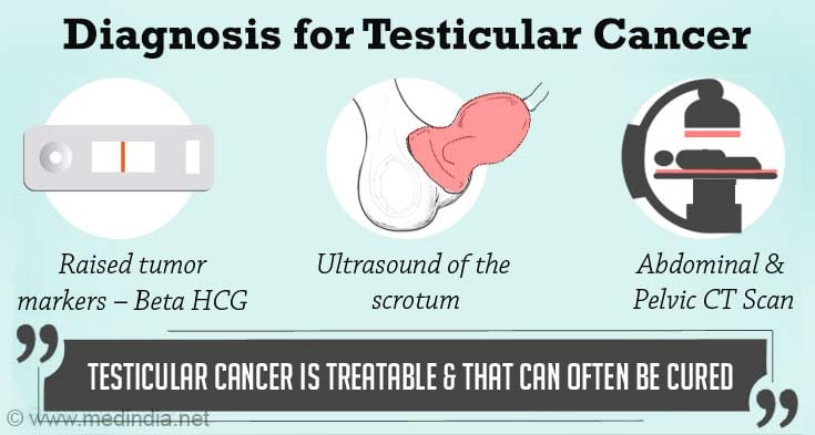
Further tests may be advised to confirm the diagnosis of testicular cancer. These include:
- Blood tests for Tumor Markers - Blood tests are carried out in the laboratory to detect and quantify tumor markers, such as AFP (Alpha Feto Protein), beta HCG and the enzyme LDH, that are specific for testicular cancer.
AFP normal value- < 16 ngm / ml
HCG normal value – <1ng/ml
- Testicular Ultrasonography- A scrotal ultrasound can detect a lump, identify its location and evaluate its size and nature.
- CT Scan- More sophisticated imaging studies, such as the CT scans of the chest; abdomen and pelvic area help to identify the extent of the cancer spread or metastases. Lesions as small as 1 mm can be detected by some of these imaging machines.
- CT Scan or MRI of the brain is a must for patients with stage III or stage IV disease.
- Chest X-ray is also done as part of the routine tests.
- Inguinal Orchiectomy is the surgical excision of the testicles along with the associated structures, such as the epididymis. A biopsy is usually not preferred as it may result in the spread of cancer cells. The resected specimen is subjected to a histological examination, by a pathologist, for differential diagnosis.
About Tumor Markers:
|


