Medindia » Multimedia » Slideshows » Eye Allergies

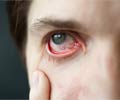
Eye allergies occur when allergens irritate the conjunctiva or the delicate membrane covering the eye and the eyelid. The body recognizes these allergens as foreign objects and triggers an immune response by releasing immunoglobulin E (IgE) molecules. This results in reactions, such as itchiness, watering eyes, soreness, swelling, among other symptoms. Eye allergies occur along with allergic rhinitis or asthma and do not cause a threat to vision.

Eye allergies are of 5 main types. They are classified as follows:

The causes of eye allergies are similar to those that cause allergic rhinitis. The allergens can be classified into 3 categories. They are as follows:

Itching (88%), Soreness in the eyes (75%), Watery eyes (88%), Stinging (65%) and Redness in the eyes (78%)
Other symptoms are swelling of the eyelid, light sensitivity, pain, eye discharge, and grittiness. Severe symptoms of eye redness are observed in conditions, such as glaucoma, keratitis, and uveitis. The symptoms of ocular allergy are severe in the case of seasonal allergies as compared with indoor allergies.
Dryness in the eyes: Dryness in the eyes occurs due to contact lens use, extended use of the computer, use of certain drugs, or old age. Diagnosis includes detecting an abnormal staining of tears, abnormal breakup time of tears, and a thin tear film.
Toxic conjunctivitis: This is observed with prolonged use of over-the-counter eye medications or eye drops. Blepharitis: This is a bacterial infection of the eyelids that results in the inflammation of the eyes.
Redness of the eye: Bacteria or viruses can cause eye redness more frequently in children. Bacterial conjunctivitis and viral conjunctivitis are less frequently observed in adults. In bacterial conjunctivitis, there is a yellow-green mucus discharge. Streptococcus pneumoniae, Haemophilus influenzae cause bacterial conjunctivitis in children, while Staphylococcus aureus cause bacterial conjunctivitis more frequently in adults. Herpes simplex viral infections can cause light sensitivity, mucus discharge, and pain. Viral infections cause burning and water discharge in the eyes.
Contact dermatoconjunctivitis: This is a familiar condition, where there is an inflammation of the eyes with the use of contact lenses, cosmetics, or eye medications.
The movement of the eye, sharp vision, and the exact presence of redness are examined. The conjunctiva or the thin layer covering the eyelid and the eye is also examined to detect bumps. Rashes in the eyelids or face are also examined to understand the type of allergy in the individual.
Blood tests: The presence of white blood cells, eosinophils, is analyzed to detect the presence of an allergy. The conjunctiva is scraped to detect the presence of eosinophils. Tear cytology, impression cytology (analyzing the epithelial cells of the conjunctiva), and brush cytology are analyzed to understand the effects of allergens on the eye.
Patch tests: These tests are used to identify allergic reactions that are not triggered by IgE. Cosmetics and other allergens are used to detect the allergic reactions. If the patch test turns out negative, then a repeated open application test (ROAT) can be performed.
Confocal microscopy: Ocular allergic inflammations can be studied and detected with confocal microscopy. Staining tests: The tear film can be stained to detect the integrity of the tear film. Lissamine green and rose bengal stain epithelial cells that are damaged or dead. Fluorescein stains defects in epithelial cells. Tear film production is assessed by the Schirmer test. The Schirmer test is widely used to assess dry eyes.
Tear IgE measurements: IgE levels in tears increase in those who have ocular allergies. Eosinophilic cationic protein (ECP) levels are increased in tears. Measuring ECP levels can specifically indicate the involvement of the cornea in VKC.
Tear-specific IgE assay: It is similar to serum-specific IgE measurements. However, it is difficult to obtain tears, dilute the tears, and quantitate the IgE levels. Inflammatory mediators: These are indicative of allergic responses in tears and measured with membrane arrays or bead-based flow cytometry.
Skin prick tests (SPTs): Detect the presence of allergens, such as pet dander, mites, molds, cockroaches, latex, and pollen.
If SPTs are unable to detect IgEs triggered by known antigens, serum-specific IgE is measured in response to specific pure allergens.In cases where both the above methods do not work conjunctival allergen challenge (CAC) or conjunctival provocation tests (CPTs) are recommended for individuals with persistent symptoms. In this case, individuals who are symptom-free and do not have inflammation in the eye, but complain of eye allergies, are injected with allergens in the conjunctiva.
Avoiding allergens: Individuals, who are affected by pollen, dust mites, and latex, among others can try to avoid contact with these allergens.
Anti-allergy medications: Medications, such as vasoconstrictors, corticosteroids, mast cell stabilizers, antihistamines, mast cell stabilizers with antihistamines (dual-action drugs), calcineurin inhibitors, and nonsteroidal anti-inflammatory drugs help suppress the symptoms. Specific immunotherapy: This treats SAC, PAC, and occasionally VKC. It is effective in reducing the allergic symptoms along with the use of eye drops. Educating the patients: This involves providing information to the patient on the condition, the cause, and the form of preventive measures to manage the disease.
Seasonal allergic conjunctivitis 9 (SAC) and PAC display IgE-mediated immune reactions. The treatment regimen involves the following:

Vernal keratoconjunctivitis (VKC) and atopic keratoconjunctivitis (AKC) display non-IgE- and IgE-mediated immune responses. The treatment regimen involves the following:

Contact blepharoconjunctivitis displays a non-IgE mediated immune response. The treatment regimen involves the following:
 MEDINDIA


