What is Myotomy?
Myotomy is a surgical procedure, which is used to divide a muscle.
A common procedure is Heller myotomy, which is done to treat a condition called achalasia cardia.

About Achalasia Cardia
Food passes from the mouth into the stomach via a muscular tube called the food pipe or the esophagus. It extends through the central chest up to the upper abdomen, where it opens into the upper end of the stomach. The lower end of the esophagus is surrounded by thickened muscle, which forms a sphincter at the junction of the esophagus and the stomach. The sphincter has two important functions, to control the passage of food into the stomach and to prevent reflux of acid up into the esophagus. A lack of tone of the lower end of the esophagus results in a condition called gastroesophageal reflux disease (GERD).
Achalasia cardia is a condition where the sphincter becomes tight. Therefore, food cannot pass easily through it resulting in difficulty in swallowing that worsens with time. Patients also experience extreme discomfort in the chest, chest pain and regurgitation of food into the mouth eventually resulting in weight loss.
How does Myotomy Help in Achalasia Cardia?
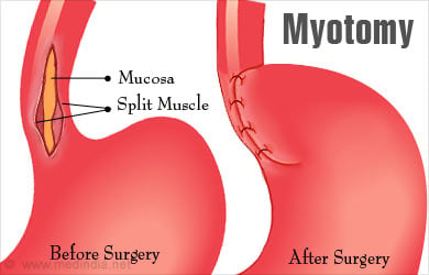
Heller myotomy is a procedure where the muscle of the lower end of the esophagus is divided longitudinally on the anterior aspect. This loosens the lower esophageal sphincter making it possible for food to pass.
What is the Procedure Adopted for Achalasia Cardia?
Heller myotomy for achalasia cardia can be done through an open surgery, or using a laparoscope or a thoracoscope. The patient is administered general anesthesia for the procedure.
- An open surgery is done through an incision in the upper abdomen. The surgery is associated with a longer time for recovery, and therefore is less preferred as compared to laparoscopic surgery.
- Laparoscopic surgery involves the use of a laparoscope, which is a tube with a camera and a light source at the end. The surgery is carried out through small incisions in the upper abdomen. Carbon dioxide gas is introduced to enable better visualization of the abdominal organs. The recovery of the patient is comparatively faster with this procedure. It may sometimes have to be converted to open procedure in case of complications like bleeding and perforation.
- Thoracoscopy is also another minimally invasive surgery where the incisions are made in the chest. However, for Heller myotomy, laparoscopic access appears to have better outcomes as compared to thoracoscopy.
During either of the above approaches, the lower end of the esophagus is identified. An incision is made above the lower end that extends to the upper part of the stomach. The incision is limited to the muscular layer without affecting the inner lining of the esophagus.
Following the procedure, since the lower esophageal sphincter becomes loose, the problem of achalasia cardia is solved. Unfortunately, acid from the stomach can reflux back into the esophagus, thereby damaging its inner lining. To prevent this, Heller myotomy is followed by a procedure called partial fundoplication. In this procedure, the upper end of the stomach is wrapped around the lower end of the esophagus partially and kept in place with sutures. This acts as a valve and controls the reflux of acid into the esophagus.
Which are the Newer Techniques Being Tried for Heller Myotomy?
There are some variations in the classic open and laparoscopic approaches to Heller myotomy, which are being evaluated for the treatment of achalasia cardia. These are:
- Laparoendoscopic Single Site Surgery (LESS): This procedure requires a single incision, which is done through the umbilical scar. It has a cosmetic benefit due to the presence of only a single scar hidden by the umbilicus.
- Robot-Assisted Myotomy: Robot-assisted myotomy is an improved surgical procedure which allows better visualization and ability to carry out the surgery. However, it is more costly as compared to laparoscopy.
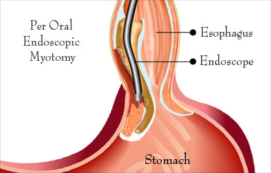
- Per Oral Endoscopic Myotomy (POEM): In this procedure, an endoscope is introduced through the mouth up to the esophagus. A tunnel is made which extends under the inner lining of the esophagus to reach the inner circularly arranged muscle of the lower esophageal sphincter, which is then cut. This procedure therefore does not have an external scar. Further experience with this procedure may increase its popularity.

What are the Complications of Heller Myotomy?
Complications of Heller myotomy include the following:
- Perforation of the esophagus, if the cut goes too deep and pierces the inner lining of the esophagus
- Incomplete cutting of the muscle, which may result in ineffectiveness of the procedure. An endoscopy at the end of the procedure can be used to confirm that the myotomy is complete
- Excessive bleeding
- Trauma to surrounding organs such as stomach, spleen and liver
- Fibrosis resulting in tightening of the esophagus
- Infection
- Too tight fundoplication or disruption of the fundoplication


