What is Percutaneous Nephrolithotomy (PCNL)?
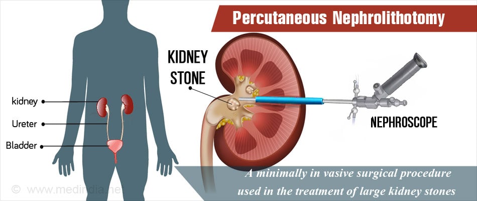
Percutaneous nephrolithotomy (PCNL) is a minimally invasive procedure used in the treatment of kidney stones bigger than 2 cm. In this procedure, a small tract (or a hole) is made through the loin leading to the kidney to carry out the surgery. A tube is inserted into the kidney through the tract created and the stone is fragmented and multiple pieces are removed through the tube.
Breaking of stone is called lithotripsy and this can be achieved using laser or pneumatic or high intensity sound energy. Various devices are available and the urologist performing the procedure may have their own preference. Percutaneous refers to the approach being made through the skin on the back (loin) to reach the kidney. Nephro means kidney and lithotomy means breaking the stone.
Why is Percutaneous Nephrolithomy (PCNL) Performed?
Percutaneous nephrolithotomy (PCNL) is done to treat kidney stones. Renal stones form due to crystallization of certain minerals / salts in the urine. Calcium oxalate stones are the most common types of urinary stones. The other urinary stones are uric acid stones, calcium phosphate, struvite (of due to infection) and rarely cysteine stones.
Percutaneous nephrolithotomy (PCNL) is the preferred method for stone removal when other methods may fail or may not be applicable due to the stone being too hard to break. Normally ESWL is used for breaking the stones in the kidney up to 2 to 2.5 cms and can be done as an out-patient procedure and it uses high intensity acoustic waves. The indications for PCNL include:
- Stones bigger than 2 cm in size
- Stones smaller than 2 cms that are too hard to break using ESWL
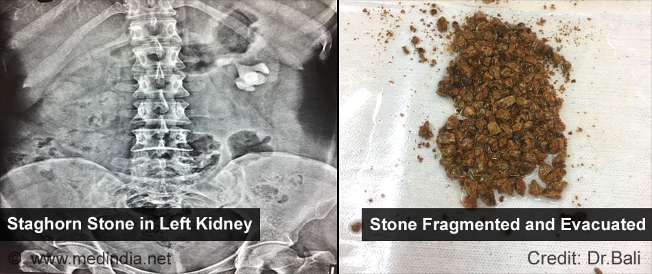
- Staghorn or calcium phosphate calculi that that have multiple horns or branches like the horn of a ‘stag’ and are distributed all over the kidney. These are highly usually difficult to remove. Struvite stones consist of magnesium aluminum phosphate that form in patients with a urinary tract infection
- Stones in the lower pole of the kidney that are not amenable to ESWL. The procedure can also be used in stones of the upper poles not responding to other treatments
- Cysteine stones that occur in patients with an inherited condition called cysteinuria, where excess cysteine appears in the urine
- Stones in kidneys that have some anatomical anomaly (like horse shoe kidney)
How should one Prepare before Percutaneous Nephrolithotomy?
One has to undergo routine tests as well as other tests to assess kidney structure and function before the procedure. Imaging tests that had diagnosed the stone earlier will be reviewed by the surgeon.
1. Routine tests: Routine tests which are done before any surgery include:
- Blood tests like hemoglobin levels, blood group, and kidney function tests
- Other blood tests – Coagulation profile, Virology status for hepatitis.
- Urine tests. Urine infection should be ruled out prior to the procedure
- Electrocardiogram (ECG) to study the electrical activity of the heart
- Chest x-ray
- Plain X-ray showing Kidney, ureter and bladder (or KUB)
In older patients, a detailed assessment of the heart may be required to make sure that they are fit for surgery.
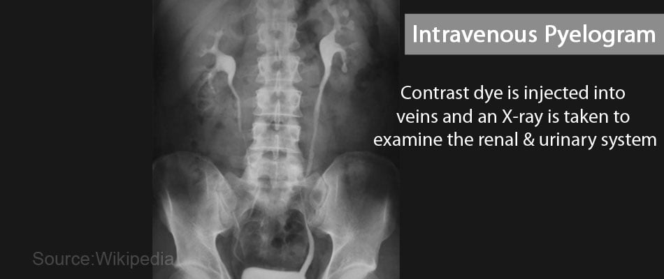
2. Assessment of kidney structure: Assessment of kidney structure or anatomy is done by using tests including intravenous pyelogram (IVP) or a CT scan.
Rarely and radioisotope (DMSA or DTPA) scans maybe required if the kidney functions are compromised.
Percutaneous nephrolithotomy should not be done if the kidney is non-functional and it may be better to remove the kidney.
- Intravenous pyelogram (IVP): During the procedure, contrast material is injected into a vein. The contrast passes through the urinary tract, during which multiple x-rays are taken at different time intervals. The test helps to understand the structure and to some extent the function of the kidney and helps localize the stone. IVP are outpatient procedures and usually requires prior preparation.
- Plain or contrast CT scan of the Kidney, Ureter and bladder – This investigations is replacing IVP as it is quick and gives a better image of the anatomy that is almost three dimensional. It also shows the location of other organs like colon, intestine, spleen and pleura in relation to the kidney. Plain CT can be done in a very short time and image reconstruction is very useful when PCNL is planned. CT scans are outpatient procedures and usually require minimum preparation.
- Radioisotope (DMSA or DTPA) scans: In these scans, a radioactive substance is injected into the vein. It passes through the kidneys and the radiation it emits can be measured with the help of a gamma camera while you lie down on the scanning table. The tests help to detect structural and functional problems of the kidneys. Radioisotope scans are outpatient procedures and usually do not require prior preparation.
How is Percutaneous Nephrolithotomy Performed?
1. Prior to the Procedure:
If you are a patient undergoing PCNL please note the following –
- Pre-operative Check-up – Routine tests as indicated above are ordered a few days before the surgery. Medications like aspirin or other blood thinners should be stopped about a week prior to the procedure. Admission is required a day before the surgery.
- The Day before the Surgery - An enema is administered the previous afternoon or evening before the surgery.
- Fasting before the Surgery - Overnight fasting is required and occasionally intravenous fluid maybe required to keep you well hydrated. Sedation is sometimes required for good overnight sleep before the surgery.
- Shift from the Ward or Room to the Waiting area in the Operating room - An hour or two before the surgery, you will be shifted to the operating room waiting area on a trolley. Once the surgical room is ready, you will be shifted to the operating room.
- Shift to the Operating room - The ambiance in the operating room can sometimes be very scary and a small amount of sedation can help in overcoming your anxiety. From the trolley, you will be shifted onto the operating table. As you look up, you will see the operating light console and at the head end will be the anesthesia machine. There may also be monitors to check oxygen levels, ECG and other vital parameters. A constant beeping sound may be present from the monitors, which may sometimes be irritating. But these are monitoring your heart beat and other vital parameters.
- Anesthesia before Surgery – The type of anesthesia depends on your preference and underlying health conditions, the surgeon’s comfort and the expected duration of the procedure.
Most of the PCNL procedures are done under general anesthesia, you will be asleep during the procedure and will not be aware of what is going on. The anesthetist will inject drugs through an intravenous line and make you inhale some gases through a mask that will put you in deep sleep. Once you are asleep, a tube will be inserted into your mouth and windpipe to administer the anesthetic gases to overcome pain and keep you comfortable while the surgery is going on.
Rarely the procedure is done under a high spinal anesthesia. You will receive an injection in your back while you sit at the edge of the table with your back bent or lie down curled up on the table. You will soon feel numbness and will be unable to move waist downwards. You will be awake during the procedure but will not be able to move your lower body waist-downwards.
2. During the procedure
Initially during the procedure, a catheter will placed through the urethra to drain out the urine. You will be placed on your stomach (or prone) on the operating table. Your joints will be cushioned to prevent pressure on them during the surgery. An antimicrobial solution will be applied to the loin. Your body will be draped and only the operative area will be left open.
A small cut will then be made on the loin and a needle will be passed up to the kidney using either an ultrasound or X-ray to show the path. Urine coming out of the needle indicates that the needle has entered the kidney space.
Animation Video to Show the Surgical Steps of Percutaneous Nephrolithotomy (PCNL)
Next a guide wire will be passed through the needle, which will then be removed, and dilators will be passed over the guide wire. A hollow straw like sheath will be placed to keep the tract open while using the last dilator. An endoscopic instrument called a nephroscope will be inserted through the hollow sheath and the stone will be located and fragmented using the preferred energy source. The fragments will be removed with the help of a grasper or through suction.
A drain or nephrostomy tube will be placed through the opening in the back. This drain and the catheter are usually removed after 24 to 48 hours. A DJ stent maybe left from the kidney to the bladder if some stones are difficult to remove and an ESWL is planned.
Surgeons often perform the procedure under ultrasound or fluoroscopic guidance and check for any residual fragments that maybe left behind before completing the procedure Once removed, the stones are sent for chemical testing.
Variations of the procedure
Variations to the above procedure of percutaneous nephrolithotomy include the following:
- Single step dilatation: A one-step balloon dilatation is sometimes done instead of using repeated dilators. The blood loss is less with this procedure. But this depends also on the anatomy of the kidney and the ease of the procedure.
- Mini-perc, ultra-mini perc and micro-perc procedures: In these procedures, smaller tracts are used and the stones are fragmented during the procedures. Ultra-mini perc and micro-perc procedures are used for smaller stones (1 cm size or less). All the fragments may not get washed out and may come out through the urine. The procedures result in reduced bleeding and pain, and earlier discharge from the hospital.
- Tubeless PCNL: Tubeless PCNL is PCNL where a drain or nephrostomy tube is not placed following the procedure. A substance like fibrin glue is inserted into the nephrostomy tract to close it
- Retrograde PCNL: In the retrograde PCNL procedure, the guide wire is passed into the kidney from the urethra with the help of an ureteroscope and then passed to the flank and the dilatation is then over the guidewire that comes from below. The procedure is a little cumbersome but the puncture is more accurate.
3. After the Procedure
- Waking up from General Anesthesia – If you have received general anesthesia, once the surgery is over, you will wake up and the tube down the wind pipe will be removed. You will be asked to open your eyes before the tube is removed. You will still be sedated a bit and the voice of the anesthetist may be faint. Once the tube is out, you may have cough and sometimes nausea.
You will still have the urinary catheter and the nephrostomy tube or drainage tube through the back. There will also be an intravenous line. You will remain on oxygen support. Once fully awake, you will be shifted on the trolley and taken to the recovery room.
- Recovery room – In the recovery room, a nurse will monitor your vitals and observe you for an hour or two before shifting you to the room or a ward.
- Post-operative recovery – You will remain in the hospital for 2 to 4 days following the procedure. You will receive intravenous fluids and will be allowed to take in liquids the next day. Plain X-rays and Ultrasound maybe done to check position of the tube and check for residual fragments if any.
- Chest physiotherapy may be started after 24 hours to prevent chest infection.
- DVT Prophylaxis – Early movement of your legs and some mobilization prevents DVT or deep vein thrombosis, where a clot is formed in the deep veins of the legs. The clot can travel up to the lungs and even be fatal. Other measures like a small dose of heparin and special stockings may also be used to prevent DVT. Pain relievers and antibiotics may be prescribed.
- Discharge from hospital –. Once your parameters are all normal and the tubes have been removed you will be discharged from the hospital. Medications like potassium citrate may be prescribed. You will also be advised measures to prevent repeated kidney stones, for example - drinking plenty of water and other fluids.
What are the Advantages and Disadvantages of PCNL?
As compared to the open surgical procedure for the removal of large kidney stones, PCNL causes lesser damage to the kidneys, and results in less post-operative pain and a quicker recovery for the patient. There is no open wound to heal. The scar is about a cm in size or even less compared to a 10 to 12 cms wound and scar in an open procedure. Complications associated with the PCNL procedure are:
- Complications due to anesthesia
- Bleeding – is the single most important complication that may require blood transfusion or re-exploration and controlling the bleeding. However such a complication happens in less than 2 to 5 % of the cases.
- Injury to the covering of the lungs called the pleura, especially when the stone is in the upper pole of the kidney and the puncture is through the ribs. As a result, air and/or fluid may accumulate in the chest resulting in pneumothorax, hydrothorax or hydro-pneumothorax, which can interfere with breathing. A chest tube to drain the air or the fluid may be necessary
- Injury to surrounding organs like enlarged liver and spleen, gall bladder, duodenum, small and large intestine, or major blood vessel resulting in bleeding
- Fever and infection
- Repeat procedure or alternate procedure may be needed if the PCNL procedure is not completely successful or if the stone recurs

